Corneal Disease of the Eye

What is the Cornea?

Just like a camera, the human eye has many distinct parts that must function together to produce clear vision. The eye converts light into an electrical signal. The optic nerve transmits that signal to the brain. The brain converts this electrical signal into an image.

One important part of the eye that focuses your vision is the cornea – the eye’s outermost layer. There are many types of disorders that can affect this vital aspect of the eye, from simple allergies to complex infections. Continue reading below to learn more about corneal diseases, and how your ophthalmologists recommend treating them.

What does the cornea do?

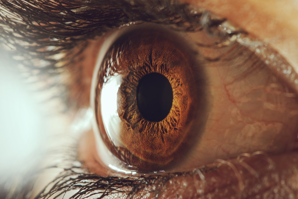
Before we dive into the different corneal diseases, we should first understand the basics of how the cornea works. When light enters the eye it initially encounters the tear film. The tear film coats the cornea, which is like the crystal clear window of the eye.

The cornea is clear, dome-shaped, and covers the front of the eye. It is made of tissues; though unlike other tissues in the body that rely on blood vessels to nourish and protect it, the cornea relies on tears and a fluid called “aqueous.” This fluid resides behind the cornea in the anterior chamber. Aqueous is responsible for maintaining eye pressure. Any trouble with aqueous production or drainage can lead to high pressure within the eye, causing glaucoma. Meanwhile, inadequate tear production can lead to dry eye syndrome.

Corneal Conditions, Disorders, & Diseases

If you suffer from seasonal eye allergies (typically caused by pollen during warm weather) consult your ophthalmologist. There are many types of medical treatments for eye allergies available by prescription. If normal allergies lead to excessive eye pain, tearing, itching, or swelling, be sure to speak with your ophthalmologist about receiving a prescription.

Some people are not able to produce enough tears or a good enough quality of tears to keep their eyes moist, healthy, and comfortable. This causes dry eye and, if symptoms persist, dry eye syndrome. Dry eye often increases with age as tear production naturally slows down. For women, this is especially true after menopause. Your ophthalmologist can diagnose dry eye through an examination of your eyes. Sometimes a tear production test will be necessary to confirm diagnosis.

This condition is typically referred to as pink eye. Conjunctivitis is an inflammation or infection of the conjunctiva – the clear outer covering of the white part of the eye. The infection makes the eye appear pink because the blood vessels of the conjunctiva become dilated. Conjunctivitis can be caused by either a bacterial or a viral infection.

This is a progressive corneal disease that affects the cornea by reducing the endothelial cell count. Because Fuchs’ Dystrophy is a progressive disease, the changes to the cornea can interfere with your vision over time. Fuchs’ Dystrophy usually occurs in people over the age of 40. To diagnose and monitor Fuchs’ Dystrophy an ophthalmologist checks for adequate endothelial cell numbers and function, obtains an endothelial cell count, and may measure the thickness of the cornea. If Fuchs’ Dystrophy causes vision loss that begins to interfere with your life, your ophthalmologist may perform a complete corneal transplant. New procedures are also available to replace only the diseased part of the cornea, thus resulting in faster rehabilitation and restoration of your vision.

A corneal dystrophy occurs when any layer or tissue within the cornea begins to weaken and break down, or when the cornea builds up a cloudy material that impairs vision. While there are many kinds of corneal dystrophies, it is a relatively rare disease and less than 1% of all cases of blindness or partial-sightedness can be traced back to it. Though there are dozens of specific corneal dystrophies, most share many common elements in that they:

  • Progress slowly
  • Are hereditary
  • Affect both eyes evenly
  • Are not typically caused by outside factors (diet, exercise, climate, or pre-existing medical conditions)

The Herpes Zoster virus usually makes its first appearance during early childhood in the form of chickenpox. Later in life the virus may reactivate, causing a rash of small blisters, which form crusts and can leave scars. When the chickenpox virus returns a second time it appears in the form of shingles. Shingles are quite painful, and have proven hard to manage with symptoms such as:

  • Pain
  • Tingling
  • Headaches
  • Numbness

If shingles affects the face it is critical that you see your ophthalmologist because it can just as easily infect your eyes. If you fear that shingles may be spreading to your eyes, your ophthalmologist can look for elevated pressure in the eye, inflammation inside the eye, and lesions on the surface of the eye. Though this corneal disease is painful, today new oral antiviral medications are providing shingles patients with a quicker, more complete recovery.

One of the main corneal dystrophies is Keratoconus, which is an uncommon condition where the cornea becomes thin and develops a cone-like bulge. As the condition progresses, the shape of the cornea is altered and vision becomes distorted. The causes of Keratoconus are unknown. Keratoconus usually begins in the teen years, but it will typically progress slowly for 10 to 20 years before the symptoms become more advanced. Keratoconus is usually corrected with eyeglasses. However, as the condition develops, rigid contact lenses may be needed to improve vision. Your ophthalmologist may recommend a corneal transplant if vision is greatly impacted. Corneal transplants offer the best prognosis for clear vision, but will not cure Keratoconus.

 Herpes of the eyes (also known as ocular herpes) is caused by the same herpes simplex virus that is largely responsible for cold sores and fever blisters. Ocular herpes is the most common infectious cause of corneal blindness in America. The disease typically causes painful sores on the eyelid or cornea and can eventually cause the cornea itself to inflame. This leads to the breakdown and destruction of cells within the cornea, causing scarring and blindness. Though the virus itself never leaves the body, ocular herpes breakouts can be controlled and treated with prescriptions from your ophthalmologist.

A pterygium is a mass of fleshy tissue that grows over the cornea. It may remain small or grow large enough to interfere with vision. The exact cause of pterygium development is not known, but statistics show that pterygiums tend to occur more frequently in people who spend a lot of time outdoors in the sun. As such, ophthalmologists believe there is a connection between pterygiums and long-term exposure to sunlight, especially to ultraviolet rays.

Protecting the eyes from excessive ultraviolet light with proper sunglasses, avoiding dry, dusty conditions, and using artificial tears are all recommended ways of treating and preventing pterygiums. If a pterygium becomes red and irritated, eyedrops or ointments can be used to help reduce the inflammation. If the pterygium grows large enough to threaten sight it will need to be surgically removed by your ophthalmologist.

Person Icon
Patient Portal
Calendar Icon
Appointments

Contact Us Now

If you live in Austin, Southwest Austin, or Kyle, TX and are in search of a great eye doctor, look no further than Howerton Eye Clinic. We are dedicated to helping our patients achieve and maintain healthy, beautiful vision. Contact us now!

South Central Austin

2610 South IH 35,
Austin, TX 78704

(512) 443-9715

Southwest Austin

5625 Eiger Rd., Suite 100,
Austin, TX 78735

(512) 872-6059

Kyle

4282 Farm to Market 1626, Suite 200,
Kyle, TX 78640

(512) 872-6064
WARNING: Internet Explorer does not support modern web standards. This site may not function correctly on this browser and is best viewed on Chrome, Firefox or Edge browsers. Learn More.